|
|
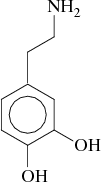
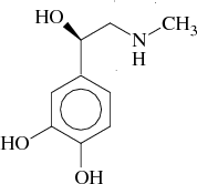
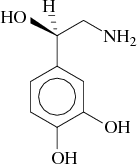
The catecholamines dopamine, L(–)-epinephrine and L(–)-norepinephrine (Figure 1.1-1.3) form a family of neurotransmitters and hormones, which mediate central and peripheral excitation as well as inhibition.
The release of catecholamines occurs in the adrenal medulla and is regulated by preganglionic neurons of the thoracic medulla (T5 – T11) via cholinergic synapses. Depolarisation of preganglionic axons in humans normally leads to a release of both, epinephrine and norepinephrine in the ratio of about 4 to 1. Under resting conditions only small amounts of epinephrine and norepinephrine are secreted from the adrenomedullary cells.
In the case of internal and external stress, caused e.g. by infections, hypoglycemia, hypoxia, hypothermia, thermal burn or pain, secretion of epinephrine and norepinephrine by the adrenal medulla is elevated. The two transmitters provide a state of increased efficiency for the organism by the following mechanisms:
Catecholamines exert their cellular action by binding to membrane proteins known as adrenergic receptors (see section 1.4). At present, several mechanisms which terminate the action of catecholamines are known [66] :
© 2001 Alexander Binder