All b1 adrenoceptors mediate responses to noradrenaline released from the sympathetic nerve terminals and to circulating adrenaline. b1 adrenoceptors were originally defined based on their rank order of potency of these endogenous agonists and by the synthetic agonist isoprenaline. Isoprenaline has shown to be the most potent of the three agonists with adrenaline and noradrenaline showing equal affinity for this particular receptor.
Subsequently, synthetic antagonists have been identified for b1 receptors these include the non-selective propranolol as well as the more selective compounds like metoprolol, practolol, atenolol and betaxolol. The selective b1-adrenoceptor agonists so far identified (denopamine, xamoterol and Ro 363) have limited utility as pharmacological tools because of low selectivity and/or efficacy.
The major ligands used for the autoradiographic localisation of b-adrenoceptor subtypes are [125I] cyanopindolol, [3H] dihydroalprenolol and [125I] pindolol. Of these [125I] cyanopindolol is the most commonly used because of its high affinity, ease of preparation, relatively short exposure time, and selectivity for b-adrenoceptors.
Catecholamines acting on b1 adrenoceptors mediate a number of tissue responses including increases in cardiac rate and force of contraction, stimulation of renin secretion, relaxation of coronary arteries and relaxation of gastrointestinal smooth muscle.
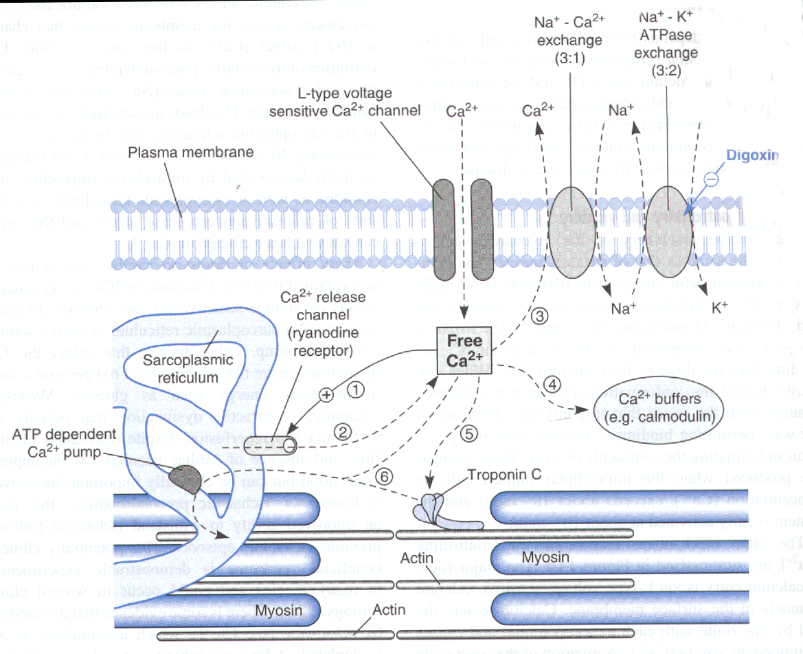
All known adrenoceptors are coupled to their effector systems by guanine nucleotide binding proteins (G proteins). b1 adrenoceptors are positively coupled to the membrane bound enzyme adenylate cyclase via activation of Gs G-protein. Activation of adenylate cyclase produces alterations in cellular activity by increasing intracellular levels of cAMP. In the myocardial cell increases in cAMP cause more L-type voltage sensitive Ca2+ channels to open in response to depolarisation. Calcium then enters the cell by this route with each action potential and causes an immediate rise in the concentration of intracellular calcium.

Diagram from Pharmacology (4th edition) Rang Dale and Ritter
Raised levels of intracellular calcium stimulate further calcium release from the sarcoplasmic reticulum (see diagram above). Activation of the contractile machinery is due partly to this influx of calcium but mainly to the secondary release from the sarcoplasmic reticulum.
The free intracellular Ca2+ binds to troponin C causing a change in conformation of the troponin complex with the result that tropomycin shifts out of the way permitting binding of myocin cross bridges to actin initiating the contractile process this leads to contraction of the myocardial cell.
Both the heart rate (chronotropic effect) and the force of contraction (inotropic effect) are increased, resulting in a markedly increased cardiac output and cardiac oxygen consumption. The cardiac efficiency is reduced. Catecholamines can also cause disturbance of the cardiac rhythm, cumulating in ventricular fibrillation.