As mentioned above, the 2ar is linked to a guanine nucleotide regulatory protein. Several types of these proteins have been described[21] (see table tab:g-table).
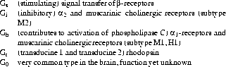
Table 1.4: Guanine Nucleotide Binding Protein Classes
The G-proteins are located at the cytoplasmic side of the cell membrane
and consist of three subunits: ![]() (39-52kDa),
(39-52kDa), ![]() (35-36kDa) and
(35-36kDa) and
![]() (8-10kDa).
The sequence of events leading to signal transduction by the
(8-10kDa).
The sequence of events leading to signal transduction by the
![]() -adrenergic receptor can be described as follows[21, 20, 34]:
-adrenergic receptor can be described as follows[21, 20, 34]:
The binding of an agonist to the binding domain of the receptor
is followed by binding of the receptor
to the ![]() -subunit of the G protein. As a consequence the G protein is
activated (substitution of GTP for GDP) and the receptor is
phosphorylated by a specific kinase. This phosphorylated receptor
dissociates from the cell membrane and translocates into the cell. The G protein
dissociates to its subunits. The
-subunit of the G protein. As a consequence the G protein is
activated (substitution of GTP for GDP) and the receptor is
phosphorylated by a specific kinase. This phosphorylated receptor
dissociates from the cell membrane and translocates into the cell. The G protein
dissociates to its subunits. The ![]() subunit, now bound to GTP,
stimulates the adenylate cyclase which is
located at the cytoplasmic side of the cell
membrane, resulting in a plasmatical cAMP concentration rise. The G
protein can also interfere in ion channel regulation. The bound GTP
is converted to
GDP and inactivates the G protein. The activity of the
adenylate cyclase decays. The receptor is dephosphorylated and re-enters the
cell membrane, eventually completing the cycle. In stage this the receptor can again
react with a ligand.
subunit, now bound to GTP,
stimulates the adenylate cyclase which is
located at the cytoplasmic side of the cell
membrane, resulting in a plasmatical cAMP concentration rise. The G
protein can also interfere in ion channel regulation. The bound GTP
is converted to
GDP and inactivates the G protein. The activity of the
adenylate cyclase decays. The receptor is dephosphorylated and re-enters the
cell membrane, eventually completing the cycle. In stage this the receptor can again
react with a ligand.
The cAMP acts as second messenger: by activating protein kinases which in turn phosphorylate various proteins. The specific cell response is determined by these phosphorylated proteins and the protein kinases being present.